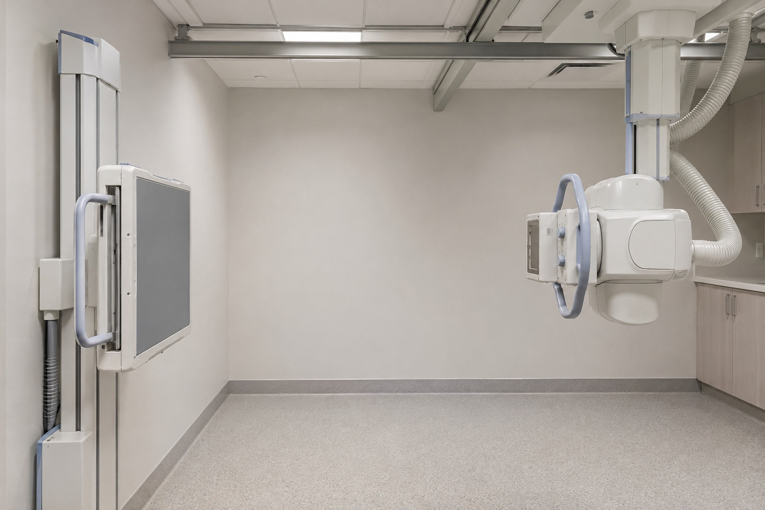
Image: AI-generated illustrative image of a radiography room. No real patient or diagnostic image is shown.
CT and MRI have transformed medicine, but a plain X-ray is still often the right place to start. The best scan is the one that answers the clinical question safely and promptly, not necessarily the most elaborate one.
X-ray, CT, and MRI: different strengths
A plain X-ray, also called a radiograph, creates a two-dimensional image with overlapping structures. It is quick, widely available, and usually less expensive than CT or MRI. Portable equipment can bring imaging to a patient who cannot safely leave their bed. These advantages matter in everyday care and emergencies. RadiologyInfo: Chest X-ray
CT also uses X-rays. A computer reconstructs many measurements into detailed cross-sectional images, reducing the problem of overlapping anatomy. It can reveal small lung abnormalities and injuries that a plain film misses. A conventional chest CT generally involves more radiation than a chest radiograph, although doses vary with the examination and protocol. CT can also be fast and lifesaving; an X-ray should not delay a CT when CT is the appropriate test. RadiologyInfo: Chest CT
MRI uses magnetic fields and radio waves, not ionizing radiation. It provides excellent detail for many soft tissues and selected heart, blood-vessel, chest-wall, and other problems. It is not a universal replacement for chest X-rays or CT: for many lung abnormalities, CT is more suitable. RadiologyInfo: Chest MRI
Common types of X-ray examinations
“X-ray” describes a family of examinations, not just a picture of a broken bone.
- Bone and joint radiographs: assess fractures, alignment, and some joint changes. Several views may be needed.
- Dental X-rays: examine teeth and surrounding bone, including decay and other dental problems.
- Chest radiographs: assess the lungs, heart outline, and surrounding chest structures.
- Mammography: specialized X-ray imaging of the breasts for screening or investigating an abnormality.
- Fluoroscopy: moving X-ray images used to assess processes such as swallowing or to guide procedures.
- CT: an X-ray-based method that produces detailed slices through the body.
The radiation exposure and preparation differ between these tests. NHS: X-ray · FDA: Medical X-ray Imaging
PA, AP, and lateral: what chest views mean
These names describe how the image is taken, not separate diseases. Both PA and AP provide a frontal view of the chest.
PA: posteroanterior, from back to front
The beam passes through the back of the chest toward a detector in front. The patient usually stands with their chest against the detector.
An upright PA image gives a useful overview of both lungs and generally allows a more reliable assessment of the heart’s apparent size than an AP image. Bringing the heart closer to the detector reduces magnification.
AP: anteroposterior, from front to back
The beam passes through the front of the chest toward a detector behind the patient. AP images are common with portable equipment when someone cannot manage a standard PA examination. The patient may be sitting, partly upright, or lying down.
AP technique can make the heart look larger than it really is. Positioning and how deeply the patient breathes also affect appearances. An apparently large heart on a portable AP image does not, by itself, prove heart enlargement. AP imaging remains valuable when it answers an urgent question safely. Radiology Masterclass: Projection
Lateral: a side view
A lateral image offers another angle. It can help locate an abnormality and reveal areas hidden on the frontal image, including behind the heart and near the diaphragm. It complements the frontal view rather than diagnosing one particular disease. Whether it is needed depends on the clinical question and local practice.
Upright and decubitus: position matters too
“Upright” describes the patient’s position, not the beam direction. “Decubitus” means lying down; a lateral decubitus examination is taken with the patient lying on one side, usually using a horizontal beam. Selected positional views can help assess fluid or air around the lungs. They are not needed for every chest examination. European Respiratory Society: Lung imaging methods
Bilateral: both sides, not a projection
Bilateral is not another beam direction like PA or AP. It means both sides. A request for bilateral knee X-rays concerns both knees. In a chest report, “bilateral changes” means changes affecting both sides; it does not name a special chest view.
What clinicians look for on a chest X-ray
The views work together. There is no simple rule that “PA reads pneumonia” while “lateral reads fluid.” Clinicians assess the whole examination and the patient’s symptoms.
Common questions include:
- Is there a lung opacity, meaning an area that looks denser than expected? Infection is one possibility, but fluid, partial lung collapse, and other causes can look similar.
- Is there a pleural effusion, or fluid around a lung?
- Is there a pneumothorax, or air around a lung that can cause it to collapse?
- Does the heart outline look enlarged, allowing for the projection and positioning?
- Are visible lines or tubes in an appropriate position?
A pneumonia-like pattern does not reliably identify the organism causing an infection. Symptoms, examination, and sometimes laboratory tests are needed. RadiologyInfo: Chest X-ray · Merck Manual: Community-acquired pneumonia
Before interpreting an apparent abnormality, the reader considers rotation, breath depth, exposure, and positioning. Technical differences can change how the lungs and heart look. This is one reason a photograph of an X-ray is not a reliable basis for self-diagnosis. European Respiratory Society: Lung imaging methods
What a normal chest X-ray cannot promise
A normal chest X-ray does not exclude every serious chest condition. Pulmonary embolism and small lung cancers may not be visible. Early pneumonia can also be missed. Persistent or worsening symptoms still need medical assessment; further tests may be appropriate even when the X-ray is reported as normal. RadiologyInfo: Chest X-ray · RCEMLearning: Community-acquired pneumonia
A normal chest X-ray also cannot rule out heart failure. The heart may require assessment with blood tests and echocardiography, depending on the symptoms and examination. Heart Failure Association of the ESC: Diagnostic consensus
Radiation: use it thoughtfully, not fearfully
X-rays use ionizing radiation, so each examination should have a clear purpose and use a dose appropriate to the task. For medically justified imaging, the expected benefit generally outweighs the small radiation risk. Tell the imaging team if you are pregnant or might be pregnant. They can choose the appropriate examination and precautions; pregnancy does not automatically mean necessary imaging must be refused. FDA: Medical X-ray Imaging · FDA: X-rays, Pregnancy and You
The takeaway: Plain X-rays, CT, and MRI are complementary tools. Understanding the views helps you understand a report, but interpreting the result requires the images, clinical context, and a trained professional.
This article provides general education, not an individual diagnosis. Seek urgent care for severe or sudden breathing difficulty or severe chest pain, even after a normal X-ray.
Sources and further reading
- RadiologyInfo: Chest X-ray
- RadiologyInfo: Chest CT
- RadiologyInfo: Chest MRI
- NHS: X-ray
- FDA: Medical X-ray Imaging
- FDA: X-rays, Pregnancy and You
- Radiology Masterclass: Chest X-ray projection
- European Respiratory Society: Lung imaging methods
- Merck Manual: Community-acquired pneumonia
- RCEMLearning: Community-acquired pneumonia
#XRay #ChestXRay #MedicalImaging #Radiology #PatientEducation #LungHealth #HealthLiteracy #EndYourIllness
