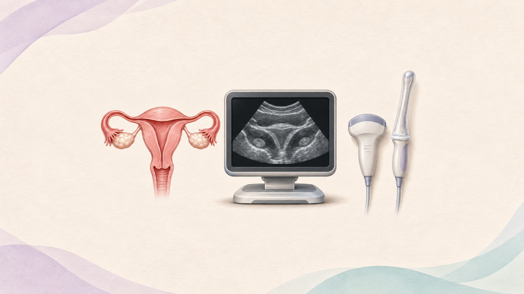
An ultrasound referral can leave you with questions: Will the scan be on my tummy or inside my vagina? What is the clinician looking for? Will it hurt?
Pelvic ultrasound helps clinicians investigate symptoms involving the womb, ovaries and nearby structures. It uses sound waves rather than ionising radiation. The best approach depends on the question being investigated, the images needed and your informed choice. ISUOG: pelvic ultrasound
Why might you need a gynaecological ultrasound?
A clinician may recommend a scan for pelvic pain, unusually heavy or irregular periods, bleeding between periods or after menopause, or a suspected lump. It can help assess the size and location of fibroids, describe ovarian cysts and examine the womb lining. The findings help decide whether observation, further tests or treatment is appropriate. RadiologyInfo: pelvic ultrasound
Scans can also support fertility investigations by examining the shape of the womb and the ovaries, including the small follicles in which eggs develop. A scan alone cannot explain every cause of difficulty conceiving or predict whether someone will become pregnant. ISUOG: pelvic ultrasound
Sometimes the purpose is follow-up: checking whether a previously identified problem has changed rather than looking for a new diagnosis. Guy’s and St Thomas’ NHS: transvaginal ultrasound
1. Transabdominal ultrasound: a scan over your lower tummy
How it is done: You lie on an examination couch. The practitioner puts gel on your lower abdomen and moves a handheld probe over the skin. No probe enters the vagina.
What it investigates: This gives an overview of the pelvis and can help assess the womb, ovaries and larger pelvic masses. An internal scan may be offered when more detail is needed.
Preparation: A full bladder is often requested because it helps the practitioner see the pelvic organs. Follow your own clinic’s instructions about how much to drink and when. Cambridge University Hospitals NHS: pelvic ultrasound
2. Transvaginal ultrasound: a closer view from inside the vagina
How it is done: After discussion and consent, a slim, covered, lubricated probe is gently placed in the vagina and moved to obtain different views. The probe does not enter the womb. You are given privacy to undress and a covering during the examination.
What it investigates: Being closer to the pelvic organs can provide clearer detail of the womb lining, womb muscle and ovaries. This is useful for investigating bleeding, pain and suspected uterine or ovarian problems.
Preparation and comfort: You usually empty your bladder first. Pressure or discomfort can occur, particularly if you already have pelvic pain. Tell the practitioner if it hurts and ask to pause or stop. A routine scan can usually be performed during a period, but a tampon must be removed. Cambridge University Hospitals NHS: pelvic ultrasound; ISUOG: pelvic ultrasound
3. Transperineal ultrasound: examining the pelvic floor externally
How it is done: A covered probe is placed against the outside of the perineal or labial area, near the vaginal opening. It is not inserted into the vagina. You may be asked to squeeze your pelvic floor or bear down so the practitioner can assess movement as well as anatomy.
What it investigates: In specialist services, this can help assess pelvic organ prolapse, certain bladder or bowel-emptying problems, and pelvic floor muscle injuries after childbirth. It answers different questions from a standard scan of the womb and ovaries, so it is not a universal replacement for transvaginal ultrasound. Bladder preparation depends on the examination. ISUOG: ultrasound in pelvic floor disorders; European Society of Urogenital Radiology: pelvic floor imaging
4. Saline infusion sonohysterography: outlining the inside of the womb
Also called SIS or a saline sonogram, this is a more specialised investigation of the womb cavity.
How it is done: A speculum is used to see the cervix. A thin tube passes through the cervix, allowing sterile saline into the womb while transvaginal ultrasound images are taken. The fluid separates the cavity’s walls so small abnormalities are easier to see.
What it investigates: It can clarify suspected polyps, fibroids projecting into the cavity, scar tissue or certain differences in womb shape, including during investigations of abnormal bleeding, infertility or repeated miscarriage.
Preparation and risks: It is commonly scheduled shortly after a period. Cramping, fluid leakage or light spotting may follow, and infection is an uncommon risk. Ask your clinic about pain relief and aftercare. It must not be performed during pregnancy or with an active pelvic infection. Tell the team if pregnancy is possible. RadiologyInfo: sonohysterography; ACOG: sonohysterography
Where do Doppler and 3D ultrasound fit?
Doppler assesses blood flow and may be added during a pelvic scan, for example when evaluating an ovarian abnormality. It is a technique, not necessarily a separate appointment. 3D ultrasound can provide additional views of anatomy, including the shape of the womb. Neither is automatically needed for everyone. A specialised contrast examination, such as HyCoSy, may be offered to investigate whether the fallopian tubes are open; an ordinary pelvic scan does not reliably answer that question. RadiologyInfo: pelvic ultrasound
What about adenomyosis and endometriosis?
Ultrasound can help diagnose adenomyosis, where tissue similar to the womb lining is found in the womb muscle. MRI may be needed if the findings are unclear or further assessment is required. NHS: adenomyosis
For endometriosis, ultrasound may show ovarian endometriomas and some deep disease. Specialist expertise matters. A normal ultrasound does not rule out endometriosis, particularly smaller or superficial deposits. Continuing symptoms deserve follow-up even when the scan looks normal. RCOG: endometriosis
Ultrasound during pregnancy
Abdominal and transvaginal scans have different roles in pregnancy. Early scans may help locate a pregnancy and assess development. Later scans assess growth, anatomy and other aspects of pregnancy care. Medical ultrasound has no known harmful effects when used appropriately, but it cannot detect every condition. NHS: ultrasound scans in pregnancy
Very early pregnancy may be too small to identify on one scan. Symptoms, repeat scans and pregnancy-hormone blood tests may be needed before the clinician can give a clear answer. An inconclusive scan does not by itself confirm miscarriage or exclude ectopic pregnancy. RCOG: bleeding or pain in early pregnancy
Your consent, privacy and choices matter
An internal scan is never compulsory simply because of your age or sexual history. Not having had penetrative vaginal sex does not automatically rule it out either. The team should discuss clinical suitability, consent and alternatives with you, with age-appropriate safeguards where relevant. British Medical Ultrasound Society: transvaginal ultrasound guidance
You can decline, ask questions or withdraw consent during the examination. Ask about a trained chaperone and tell the clinic about accessibility needs, previous difficult examinations or anything that would help you feel safer. You do not need to justify wanting privacy. If you prefer an abdominal scan, discuss what it may show and what detail might be missed. Guy’s and St Thomas’ NHS: transvaginal ultrasound
What a scan cannot promise
Ultrasound findings need to be interpreted alongside your symptoms and other tests. For abnormal bleeding, a clinician may still recommend hysteroscopy, which uses a camera to inspect the womb, or a biopsy to examine tissue. Ultrasound cannot replace those tests when they are needed. NICE: heavy menstrual bleeding
Investigating symptoms or following a known cyst is different from screening people who feel well. Routine ultrasound is not a recommended ovarian-cancer screening test for people without symptoms at average risk. ACOG: ovarian cancer
Before leaving, ask who will explain the results, when to expect them and what happens if symptoms persist.
When not to wait for a routine scan
Seek emergency care for sudden severe abdominal or pelvic pain, collapse, fainting, or heavy bleeding with marked weakness or dizziness. If pregnancy is possible, pain or bleeding needs prompt medical assessment, particularly with one-sided pain or shoulder-tip pain. Ectopic pregnancy can be life-threatening, even before you have a positive pregnancy test. In the UK, call 999 for severe symptoms or collapse; elsewhere, use your local emergency number. NHS: ectopic pregnancy symptoms; RCOG: bleeding or pain in early pregnancy
This article provides general health information and does not replace assessment or advice from your healthcare professional.
Hashtags
#WomensHealth #PelvicUltrasound #Gynaecology #Ultrasound #TransvaginalUltrasound #Fibroids #EndometriosisAwareness #Adenomyosis #PelvicHealth #PatientEducation #InformedConsent #EndYourIllness
Sources and further reading
- ISUOG: pelvic ultrasound
- RadiologyInfo: pelvic ultrasound
- Guy’s and St Thomas’ NHS: transvaginal ultrasound
- Cambridge University Hospitals NHS: pelvic ultrasound
- ISUOG: ultrasound in pelvic floor disorders
- European Society of Urogenital Radiology: pelvic floor imaging
- RadiologyInfo: sonohysterography
- ACOG: sonohysterography
- NHS: adenomyosis
- RCOG: endometriosis
- NHS: ultrasound scans in pregnancy
- RCOG: bleeding or pain in early pregnancy
- British Medical Ultrasound Society: transvaginal ultrasound guidance
- NICE: heavy menstrual bleeding
- ACOG: ovarian cancer
- NHS: ectopic pregnancy symptoms
