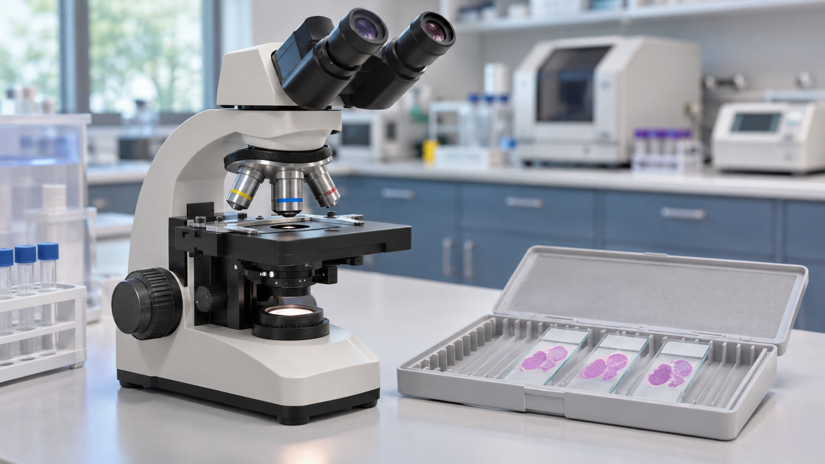
Image: AI-generated illustration of a pathology laboratory. Examining cells and tissue is a key part of diagnosing many cancers.
A lump, an abnormal scan or an unusual blood result can raise concern about cancer, but it is not automatically a cancer diagnosis. Doctors bring together the person’s symptoms, examination and test results to work out what is happening.
For most solid cancers, examining a sample of cells or tissue is the key step in confirming the diagnosis. Some cancers follow different pathways. There is no single test that reliably detects, confirms or rules out every cancer in every person. The tests needed depend on the suspected cancer and the individual situation. National Cancer Institute: How Cancer Is Diagnosed.
Screening and diagnosis have different purposes
Screening looks for signs of cancer or precancer in people without symptoms. Examples include screening mammograms and cervical screening. Diagnostic testing investigates a symptom, an examination finding or an abnormal screening result.
An abnormal screening result usually needs follow-up before anyone can say whether cancer is present. A normal screening result also does not explain a new or persistent symptom. Screening can miss disease, so symptoms still deserve medical assessment. National Cancer Institute: Cancer Screening Overview.
The starting point is a medical history and examination
A clinician asks when symptoms began, whether they are changing, and about previous illnesses, medicines, tobacco use and family history. The examination focuses on the affected area and may include checking lymph nodes or looking for other signs of illness.
Symptoms that warrant assessment include an unexplained lump, unusual bleeding, a changing mole, unexplained weight loss, or a cough or change in bowel habits that persists. These symptoms often have causes other than cancer. Cancer can also be painless, so waiting for pain is not a reliable way to decide whether to seek help. National Cancer Institute: Symptoms of Cancer.
Blood and other laboratory tests provide clues
Tests may include a complete blood count, which measures blood cells, and blood chemistry tests, which help assess organs such as the liver and kidneys. Urine or other body-fluid tests may be appropriate for particular symptoms.
Normal routine blood results do not rule out cancer, and abnormal results do not necessarily mean cancer. They need to be interpreted alongside the rest of the assessment. National Cancer Institute: Diagnostic Laboratory Tests.
Tumor markers are not a universal cancer check
Blood tumor markers, such as PSA, CA-125, CEA or AFP, can be useful in particular situations. They may contribute to diagnosis, treatment decisions or monitoring, depending on the marker and cancer type.
However, noncancerous conditions can raise a marker, and some cancers do not raise the expected marker. Ordering a broad panel of tumor markers is not a dependable way to check for every cancer. National Cancer Institute: Tumor Markers.
Imaging helps locate and assess an abnormality
Doctors choose imaging according to the body area and the question being asked. Common options include:
- Ultrasound, which uses sound waves.
- X-rays and CT scans, which use X-rays to show structures inside the body.
- MRI, which uses a magnetic field and radio waves to produce detailed images.
- PET-CT, which combines CT images with information from a radioactive tracer about activity in tissues.
Scans can help locate a suspicious area, measure it and assess possible spread. Ultrasound, CT and MRI each show different kinds of detail. National Cancer Institute: Liver Cancer Diagnostic Imaging.
A scan described as “suspicious” does not necessarily prove cancer. For example, inflammation can also take up a PET tracer. PET-CT is useful for selected clinical questions, rather than being a required test for everyone. RadiologyInfo, from the ACR and RSNA: PET-CT.
A biopsy or cell sample often provides the answer
A biopsy removes tissue for examination. Cytology examines individual cells, often collected through a fine needle or from a body fluid. Tissue samples also show how cells are arranged, which can matter when identifying and classifying a cancer.
Depending on the location, sampling may involve:
- A core needle biopsy, which removes small pieces of tissue.
- Fine-needle aspiration, which collects cells or fluid.
- An endoscopic biopsy, taken while a clinician examines an internal area with a thin viewing instrument, such as during a colonoscopy or bronchoscopy.
- A surgical biopsy, which removes part or all of an abnormal area.
- A bone marrow aspiration and biopsy, used for certain blood and bone marrow disorders.
The team chooses the procedure that can safely provide the information needed. Not everyone needs surgery, and not every patient needs every type of sample. American Cancer Society: Biopsy and Cytology Tests.
What the pathologist looks for
A pathologist is a doctor trained to diagnose disease by studying cells and tissues. In the laboratory, tissue is prepared, cut into thin sections and stained so its features can be examined under a microscope.
The pathology report may describe:
- Whether cancer is present in the sample.
- The cancer type and, when established, its subtype.
- Whether it has invaded surrounding tissue in the specimen.
- Its grade, when applicable.
- Whether cancer is present at the edges of surgically removed tissue or in sampled lymph nodes.
A small biopsy cannot necessarily answer every question about the whole tumor or its spread. Some findings become available only after additional tests or surgery. The report should be discussed with the treating clinician, rather than interpreted from one unfamiliar word. National Cancer Institute: Pathology Reports.
Special tests can identify the exact cancer type
The first microscope examination sometimes needs support from additional tests.
Immunohistochemistry, or IHC, uses antibodies to highlight particular markers in tissue. The pattern helps distinguish cancers that may look similar under the microscope. National Cancer Institute: Immunohistochemistry.
Flow cytometry measures characteristics of cells, including their markers. It is particularly useful for classifying blood cancers. Chromosome and molecular tests can identify changes in cancer cells that help establish a subtype and guide treatment. For example, the workup for acute myeloid leukemia combines blood and bone marrow assessment with specialized cell and genetic testing. National Cancer Institute: Acute Myeloid Leukemia.
Treatment biomarker testing looks for features that may make a cancer more likely to respond to certain medicines. This may involve testing proteins, genes or other markers in tumor tissue, and sometimes in blood. It is different from testing whether someone inherited a gene change that increases their future cancer risk. Neither finding an inherited risk variant nor having a family history establishes a current cancer diagnosis. National Cancer Institute: Biomarker Testing for Cancer Treatment.
Important exceptions to the usual biopsy pathway
Some leukemias can be diagnosed using blood testing. For example, chronic lymphocytic leukemia is often identified through blood counts, cell appearance and flow cytometry. A bone marrow biopsy is usually not required to establish that diagnosis. Other leukemias commonly need bone marrow testing. National Cancer Institute: Chronic Lymphocytic Leukemia Diagnostic Evaluation.
Selected liver cancers can be diagnosed through imaging. In appropriate high-risk patients, hepatocellular carcinoma can sometimes be confirmed by characteristic findings on properly performed contrast-enhanced CT or MRI without a biopsy. This requires specific criteria and specialist interpretation. It does not mean that an ordinary scan can definitively identify every liver lump or every cancer. American Association for the Study of Liver Diseases: Hepatocellular Carcinoma Guidance.
Where liquid biopsy fits
A liquid biopsy may look for tumor-derived DNA or other cancer-related material in blood. In selected patients, it can help identify molecular changes relevant to treatment, especially when tissue testing is difficult.
It does not automatically replace tissue examination or the standard diagnostic workup. A negative result cannot exclude every cancer. Blood tests marketed for detecting multiple cancers also have limitations and are not a substitute for recommended screening or investigating symptoms. National Cancer Institute: Tumor Markers and Multi-Cancer Detection Tests; ESMO: Recommendations on Circulating Tumor DNA Testing.
Staging answers how far the cancer has spread
Once cancer is identified, the team determines its extent. Some staging tests happen during the diagnostic workup. They may include imaging, examination, laboratory tests and sampling of relevant lymph nodes or other sites.
Many solid cancers use the TNM system: T describes the main tumor, N describes regional lymph node involvement, and M describes distant spread. Blood cancers and some other cancers use different systems. National Cancer Institute: Cancer Staging.
Grade and stage are different. Grade describes how abnormal the cancer looks under the microscope and helps estimate its behavior. Stage describes its extent in the body. National Cancer Institute: Tumor Grade.
What a final diagnosis means
A final diagnosis is the team’s conclusion after interpreting the relevant evidence together. For many cancers, the pathology report provides confirmation, while further reports clarify the subtype, biomarkers and stage needed for treatment planning.
Sometimes a sample is too small or the findings are inconclusive. More testing or a repeat biopsy may be needed. “Inconclusive” does not mean either “cancer” or “all clear.” Ask what is still uncertain and how the team will resolve it. American Cancer Society: Understanding Your Pathology Report.
Questions to ask while waiting for results
- What is this test intended to find out?
- Is cancer confirmed, suspected or still uncertain?
- Are special stains, molecular tests or a specialist review still pending?
- Is there enough information to establish the type and stage?
- When should I expect results, and who should I contact if they do not arrive?
- What symptoms should prompt earlier medical attention?
Results can take longer because samples need extra preparation, specialized testing or a second pathology opinion. The waiting time alone does not tell you whether a result will be good or bad. Ask for the expected timeframe for your particular tests. American Cancer Society: Waiting for Biopsy or Cytology Results.
The essential point: Cancer diagnosis is a process of answering specific questions with the right tests. A suspicious symptom or scan starts an investigation. Pathology, specialized laboratory testing and appropriate imaging help the medical team establish what is present and plan the next step.
This article provides general health information. It cannot diagnose an individual condition or replace assessment by a qualified healthcare professional. Seek medical advice for new, persistent or worsening symptoms.
#CancerDiagnosis #CancerTests #Biopsy #Pathology #CancerAwareness #PatientEducation #HealthReports #EndYourIllness
