Your doctor writes “TMT” or “2D echo” on a prescription, and suddenly you are looking at a list of unfamiliar tests. What do they mean? Are they checking for a blockage, a weak heart or an irregular heartbeat?
This Understand Your Reports guide explains the main heart tests and why your doctor might choose them. Although these reports may sit alongside your blood reports, TMT, ECG and 2D echo are cardiology tests, not blood tests. Each answers a different question.
Get urgent help for warning symptoms. New or persistent chest pressure, pain spreading to the arm or jaw, severe breathlessness, or collapse needs emergency assessment. Call your local emergency number and do not drive yourself. Do not wait for a booked test or rely on an earlier normal result. NHS: heart attack warning signs
The quick guide
- ECG or EKG: records the heart’s electrical activity, including its rate and rhythm.
- TMT: checks how the heart responds to exercise, usually with ECG and blood-pressure monitoring on a treadmill.
- 2D echo: uses ultrasound to examine the heart’s structure, valves and pumping function.
- Troponin and BNP/NT-proBNP: blood tests that help answer specific questions about heart-muscle injury or possible heart failure.
What is a TMT test called in cardiology?
TMT means treadmill test. You may also see exercise stress test, exercise ECG, treadmill stress test or exercise tolerance test (ETT). “Stress” here means the extra workload of exercise.
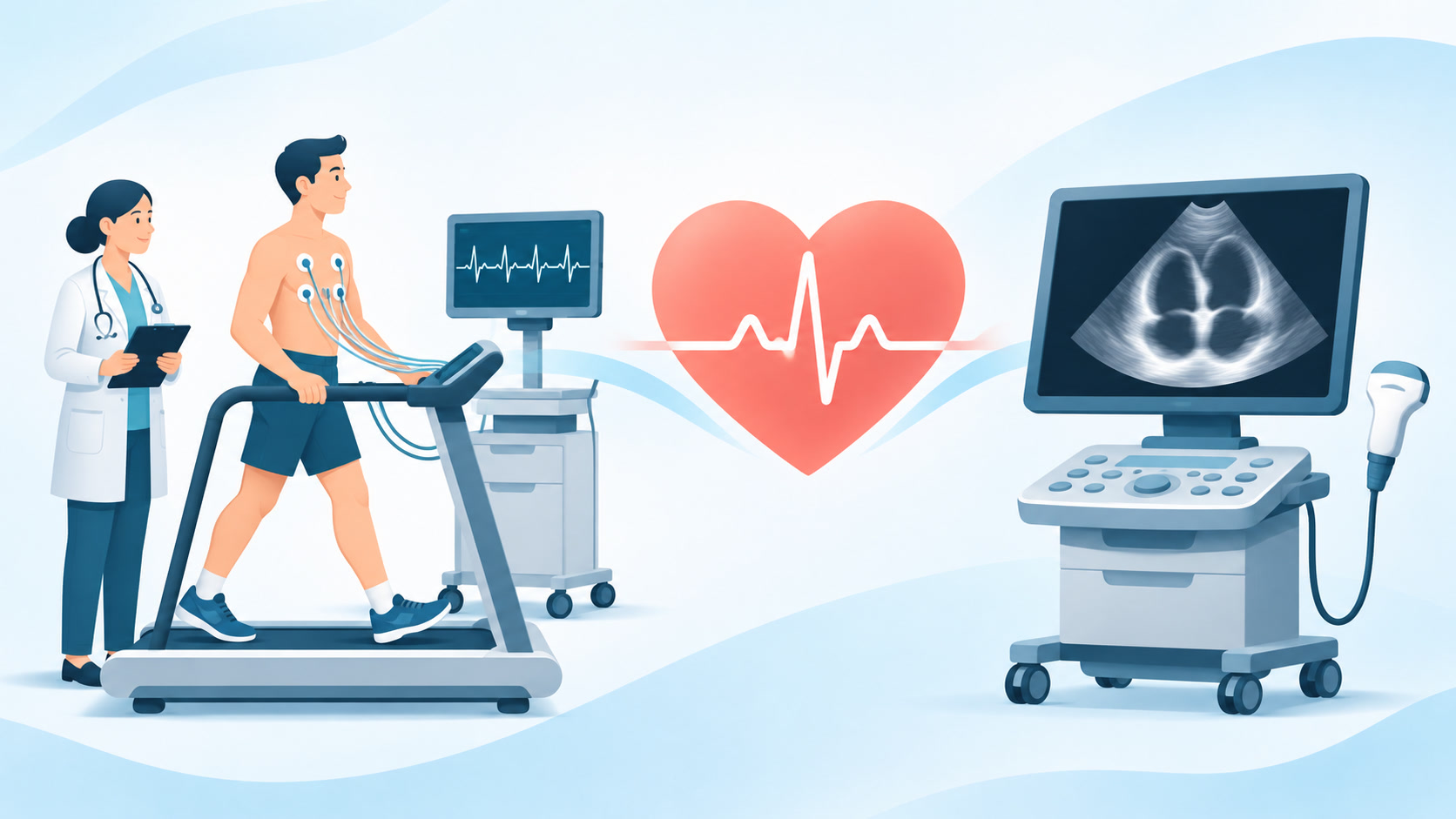
For a standard TMT, sticky electrodes record your ECG while you walk on a treadmill. Its speed and slope gradually increase. The team checks your symptoms, heart rate, rhythm and blood pressure, then monitors you during recovery. American Heart Association: exercise stress test
Why would your doctor order a TMT?
In selected patients whose condition is stable, it can help the doctor:
- Investigate symptoms brought on by exertion, such as chest discomfort or breathlessness.
- Look for ECG changes suggesting ischaemia, meaning the heart muscle may not be getting enough blood and oxygen during exercise.
- Check for exercise-related rhythm changes and assess exercise capacity.
It is most useful when you can exercise safely and your resting ECG allows meaningful interpretation. The choice depends on your symptoms, medical history and risk assessment. Some people need imaging or another test instead; others do not need further testing. TMT is not a routine requirement for everyone and is not the test used to diagnose an ongoing heart attack. AHA/ACC chest pain guideline
What can the TMT report tell you?
Your doctor considers how much exercise you managed, any symptoms, ECG changes and the heart-rate and blood-pressure response together.
- Negative for inducible ischaemia: the test did not find the expected evidence of reduced blood supply at the workload achieved. It does not rule out every form of coronary artery disease or guarantee that a future heart attack cannot happen.
- Positive or abnormal: changes need clinical interpretation. They do not, by themselves, prove that a coronary artery is blocked.
- Inconclusive or non-diagnostic: the test did not provide a reliable answer, perhaps because exercise was insufficient or the ECG was difficult to interpret.
False-positive and false-negative results can occur. Persistent symptoms may need further assessment even after a normal result. Stress-test limitations and follow-up testing
Preparation and safety
Wear comfortable clothes and walking shoes. Follow the testing centre’s instructions about food, caffeine, smoking and medicines. Tell the team about your medicines, symptoms and any difficulty walking. Do not stop a prescribed medicine unless your clinician tells you to.
TMT is supervised, and you should report chest pain, dizziness or unusual breathlessness immediately. Staff can stop the test when needed. Serious complications, including dangerous rhythm disturbances or a heart attack, are rare, but suitability must be checked beforehand. MedlinePlus: exercise stress test
What is a 2D echo?
2D echo means two-dimensional echocardiography. It produces moving ultrasound pictures of your heart. The usual chest-surface scan is called a transthoracic echocardiogram (TTE). “2D” describes the pictures; “transthoracic” describes how the scan is performed. A complete echo commonly also uses Doppler ultrasound to assess blood flow.
The sonographer moves a gel-covered probe over your chest while you lie on an examination couch. A standard resting TTE does not use ionising radiation and usually needs little preparation. Follow your appointment instructions, especially if the scan is a stress echo or a more specialised type. NHS: echocardiogram
What does it check?
An echo helps assess:
- The size of the heart’s chambers and the thickness and movement of its walls.
- How effectively the heart pumps blood.
- Whether valves are narrowed (stenosis) or leaking backwards (regurgitation).
- Certain structural problems and fluid around the heart.
Your doctor may request it for a heart murmur, suspected heart failure, valve disease, unexplained breathlessness, or follow-up of a known heart condition. It may also help assess damage after a heart attack. American Heart Association: echocardiogram
A routine resting echo does not directly map all the coronary arteries or exclude every blockage. It answers different questions from a stress test or coronary artery scan.
What does EF or LVEF mean?
Ejection fraction (EF) is the percentage of blood pumped out of a ventricle with each contraction. LVEF refers to the left ventricle, the heart’s main pumping chamber.
An EF of 60% means that around 60% of the blood in that chamber is pushed out with each beat. It does not mean that your heart is only “60% healthy”.
The American Heart Association describes a normal EF as about 55%–70%, but interpretation also depends on the measurement and clinical context. A normal EF does not exclude heart failure. In heart failure with preserved EF, the heart may have difficulty relaxing and filling even though the percentage pumped out is preserved. AHA: understanding ejection fraction
What is an ECG, and why might you need one?
An electrocardiogram, shortened to ECG or EKG, records electrical signals through electrodes on the skin. It can show heart rate, rhythm and electrical patterns that may need investigation.
Doctors use it for symptoms such as palpitations or chest pain, to investigate suspected heart disease, and sometimes to monitor medicines. A resting ECG is a short recording, so intermittent problems may require a wearable monitor. ECG and echo are different tests: an ECG records electrical activity; an echo creates ultrasound pictures. NHS: electrocardiogram
Is a stress echo the same as a TMT?
No. A plain TMT records the heart’s response to exercise without ultrasound pictures. A stress echocardiogram adds ultrasound imaging, comparing heart-muscle function at rest and under stress.
Other stress imaging tests include nuclear perfusion scans and stress cardiac MRI. A nuclear scan uses a small amount of radioactive tracer; it differs from an ultrasound echo.
If you cannot exercise adequately, the doctor may choose a medicine-based stress imaging test or another suitable investigation. Tell the team about mobility problems, asthma, pregnancy or possible pregnancy, and your medicines so they can choose safely. These alternatives have their own preparation and risk considerations. MedlinePlus: types of stress tests
Which heart tests are actually blood tests?
Troponin: checking for heart-muscle injury
Troponin I or T is measured in blood, commonly with a high-sensitivity assay. Doctors use it when assessing a possible heart attack, alongside symptoms, examination and ECG findings.
An elevated result indicates heart-muscle injury, but a heart attack is not the only possible cause. Repeat samples may be needed to see whether the level changes. A single early normal result should not be used on its own to dismiss concerning symptoms. MedlinePlus: troponin test
BNP or NT-proBNP: helping assess possible heart failure
These blood markers help doctors evaluate symptoms such as breathlessness or swelling when heart failure is suspected. They may also help monitor an established condition.
The result needs context: age, kidney function, body weight and other illnesses can affect levels. A raised value does not establish the diagnosis by itself, and lower levels do not always exclude it. Your clinician may combine the result with an echo and other findings. MedlinePlus: BNP and NT-proBNP
Five useful questions to ask your doctor
- What question are we trying to answer with this test?
- Why is this test the right choice for my symptoms?
- Should I change anything about food, caffeine or medicines beforehand?
- What does my result show, and what does it not rule out?
- What happens next if symptoms continue or the result is unclear?
Remember: a heart report is one part of your assessment. Understanding the purpose of each test helps you discuss the findings without treating one number, ECG label or “normal” result as the whole diagnosis.
This article provides general health education and cannot diagnose your symptoms or replace advice from your treating clinician.
Sources and further reading
- American Heart Association: Exercise Stress Test
- AHA/ACC: 2021 Chest Pain Guideline slide set
- MedlinePlus: Exercise stress test
- CMS: Non-emergent outpatient stress testing, including test limitations
- NHS: Echocardiogram
- American Heart Association: Echocardiogram
- American Heart Association: Ejection Fraction Heart Failure Measurement
- NHS: Electrocardiogram
- MedlinePlus: Stress Tests
- MedlinePlus: Troponin Test
- MedlinePlus: Natriuretic Peptide Tests
- NHS: Heart attack
Hashtags
#UnderstandYourReports #HeartTests #TMTTest #2DEcho #ECG #Cardiology #HeartHealth #HealthLiteracy #EndYourIllness
