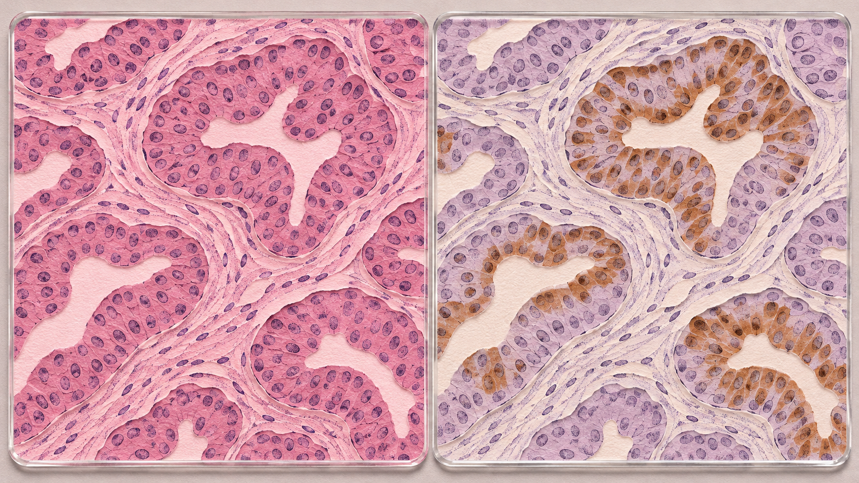
The image above is illustrative artwork, not a diagnostic tissue slide.
A cancer pathology report can contain a bewildering mix of words, percentages and abbreviations: morphology, ER, PR, HER2, Ki-67, TTF-1, positive, negative, equivocal.
These entries answer different questions. Some help identify the kind of cells in a sample. Others help describe the cancer or guide treatment. Understanding their purpose makes the report easier to discuss with your care team.
The central point is simple: pathologists interpret the appearance of the cells, their arrangement, the staining results and the clinical history together. An isolated marker result is not a complete diagnosis.
First, what is a pathology report?
A pathologist is a doctor trained to diagnose disease by examining cells and tissues. Their report records what they found in a specimen collected through a biopsy, operation or another sampling procedure.
Read the final diagnosis and any comments or later addenda before trying to decode individual stains. The report may also describe the specimen, the microscopic findings, margins, lymph nodes and additional tests. Which details can be reported depends on what was collected. An inconclusive result may lead to further testing or another sample. [1]
Morphology: what the cells look like
Morphology means the cells’ appearance and structure. Much of the initial examination uses a routine stain called H&E, short for hematoxylin and eosin. It makes cell nuclei appear blue-purple and many surrounding structures pink.
The pathologist considers both individual cells and the wider tissue pattern:
- Cell details: size, shape, the appearance of the nucleus and evidence of cells dividing.
- Tissue architecture: whether cells form glands, sheets, nests or other patterns, and how those patterns relate to surrounding tissue.
- Invasion: whether abnormal cells have grown into nearby supporting tissue, where the sample allows this to be assessed.
These features guide the choice of additional tests. IHC adds information to this microscopic assessment rather than replacing it. [2]
Grade and stage answer different questions
Grade describes features of the cancer cells and tissue that help indicate how the cancer may behave. Depending on the cancer, grading may consider how closely the tumor resembles normal tissue, cell division and other features. “Well differentiated” generally means that recognizable features of the original tissue remain; “poorly differentiated” means fewer of those features remain. Grading systems vary between cancers. [3]
Stage describes the extent of the cancer, including tumor size or local involvement, lymph nodes and distant spread. Imaging, examination, surgery and pathology may all contribute. Some staging systems also incorporate grade and biomarkers. A high grade does not automatically mean a late stage, and an IHC result alone cannot establish the complete stage. [4]
What is immunohistochemistry, or IHC?
IHC is a laboratory method that uses antibodies to detect particular proteins or other targets in a tissue sample. A visible staining reaction shows the pathologist where the target is present. Brown staining is common, but other colors and detection methods are used. [5]
The laboratory prepares thin sections, applies the selected antibodies and detection reagents, and checks the staining. The pathologist then examines the result in the relevant cells.
Think of IHC as a set of carefully chosen labels on a tissue map. The labels can help answer questions such as:
- What family of cells does this tumor belong to?
- Does the pattern support a particular cancer type or likely site of origin?
- Is a protein present that may help guide treatment?
- Is there a finding that calls for another laboratory test?
The choice of stains depends on the diagnostic question. A focused group of markers, called a panel, is often more useful than one result. Positive staining means the target was detected under that test’s conditions; it does not, by itself, mean that a cell is cancerous. [6]
Why the specimen matters
A core biopsy preserves small pieces of tissue, including some architecture. A larger surgical specimen may allow more extensive assessment of the tumor, its surroundings and surgical margins.
Cytology examines individual cells or small groups of cells, for example from a needle aspiration or body fluid. It can provide valuable diagnostic information, but usually shows less of the original tissue architecture. A cytology “cell block” can sometimes support IHC and other tests when enough suitable material is present. The laboratory needs methods validated for that preparation. [7], [8]
A small sample may not capture every part of a tumor. There may also be too few viable tumor cells for a particular test. “Insufficient material” or “not evaluable” describes a testing limitation, not a negative result. Ask whether the existing sample can answer the clinical question or whether another sample would be useful.
Reading the language of an IHC result
Depending on the marker, a report may describe:
- Positive or negative: whether the relevant staining meets that test’s interpretation criteria.
- Percentage: the proportion of specified cells showing the required staining. The denominator matters.
- Intensity: how strong the staining is, such as weak, moderate or strong.
- Distribution: whether staining is focal, patchy or widespread.
- Location: whether staining is in the nucleus, cell membrane or cytoplasm.
- A formal score: a system developed for a particular marker, cancer and clinical use.
- Equivocal or indeterminate: a result that cannot yet be assigned a clear category and may need more work.
A number is meaningful only with its marker, scoring system and tissue context. For example, “80% positive” does not mean that 80% of the person’s body contains cancer. Scores used for one marker cannot be transferred to another. [9]
Common markers, explained with their limits
The following are examples, not a checklist that every patient needs.
ER and PR: hormone receptors
ER is estrogen receptor. PR, also written PgR, is progesterone receptor. These proteins are especially important in breast cancer assessment.
For invasive breast cancer, ASCO/CAP guidance classifies ER staining in at least 1% of tumor-cell nuclei as positive. ER staining in 1–10% is reported as “ER low positive.” Evidence for endocrine treatment benefit in this group is more limited than for strongly ER-positive disease, so the comment accompanying the result matters. PR provides additional information, particularly in ER-positive breast cancer. [10]
These are breast-cancer-specific interpretation rules. An ER or PR result in another organ needs its own context. A positive receptor result can support treatment planning; it cannot guarantee that a particular treatment will work.
HER2: protein staining and gene testing
HER2 is a growth-related protein. In breast cancer, IHC usually produces a score of 0, 1+, 2+ or 3+.
- 3+ meets the IHC criteria for HER2 positivity.
- 2+ is equivocal and needs further assessment, usually in situ hybridization (ISH), which examines HER2 gene amplification.
- 0 or 1+, and 2+ with a negative ISH result, do not meet the conventional criteria for HER2 overexpression/amplification. [11]
The exact score still matters. Some treatments are used in selected advanced breast cancers with lower HER2 expression, depending on the treatment indication and other clinical findings. “HER2-negative” should therefore not be read as a permanent statement that every HER2-directed treatment is irrelevant. Ask what the exact IHC and ISH results mean for the current situation.
HER2 scoring rules can differ by cancer type. Do not apply breast scoring rules to a stomach or other cancer report.
Ki-67: a measure of proliferation
Ki-67 is associated with cells in the active cell cycle. Its percentage estimates how many assessed tumor cells show the marker.
A higher value can indicate greater proliferative activity within the relevant cancer type, but there is no single universal “high” or “low” cutoff for all cancers. Sampling, laboratory methods and how the cells are counted affect the result. Breast cancer guidance highlights these limitations. [12]
Ki-67 is not a direct measurement of how fast a particular tumor will enlarge. It should not be used alone to predict survival or decide on chemotherapy.
TTF-1: a clue to cell origin
TTF-1 means thyroid transcription factor 1. It is a nuclear protein associated with certain lung and thyroid cells. In an appropriate panel, staining can support a lung or thyroid origin. [13]
Despite its name, a positive TTF-1 result does not automatically mean thyroid cancer. A negative result does not exclude every lung cancer. The tumor’s appearance, other stains and clinical findings determine how much weight to give it.
CK7, CK20 and p40: patterns that help narrow the possibilities
CK7 and CK20 are cytokeratins, proteins associated with epithelial cells. Patterns of expression can help narrow the possible origin of a carcinoma, but many cancers share these patterns.
p40 can support squamous differentiation in the right setting. It does not identify the organ of origin on its own. Pathologists may use these stains alongside other markers when distinguishing lung cancers or considering a cancer that has spread from elsewhere. [14]
CD20 and CD3: identifying lymphocyte populations
CD20 is commonly associated with B lymphocytes, while CD3 is associated with T lymphocytes. They help characterize lymphoid cells and investigate possible lymphoma. [15]
Normal immune cells can also show these markers. Their presence alone does not diagnose lymphoma or determine its subtype. The distribution of the cells, their morphology and additional tests matter.
PSA and PAX8: further clues about origin
PSA staining examines prostate-specific antigen in tissue. It can help assess possible prostate origin in the appropriate setting. This is different from a blood PSA measurement. [16]
PAX8 can support certain kidney, thyroid or Müllerian origins, including particular ovarian, fallopian-tube and uterine tumors. It is another panel marker, not a unique label for one cancer. [17]
Mismatch-repair proteins: MLH1, PMS2, MSH2 and MSH6
These proteins help cells repair certain DNA errors. IHC can assess whether their expression is retained or lost in tumor cells.
Loss of one or more proteins, with appropriate control staining, can indicate deficient mismatch repair, often abbreviated dMMR. This may guide further tumor testing, treatment discussions or assessment for an inherited condition such as Lynch syndrome. [18]
An abnormal tumor stain does not by itself diagnose Lynch syndrome. Some changes occur only in the tumor. Follow-up may include tests such as MLH1 promoter methylation analysis or germline testing, chosen according to the cancer and staining pattern. Retained protein expression also does not guarantee that every aspect of DNA repair is functioning normally. [19]
p16: useful only in the right setting
p16 can serve as a surrogate, or indirect marker, for high-risk HPV-associated disease in particular settings, especially oropharyngeal squamous cell carcinoma. It is not universal proof of HPV infection or HPV-driven cancer in every organ. Current guidance calls for HPV-specific testing in certain circumstances. [20]
PD-L1: a treatment-related marker with different scores
PD-L1 testing may help select immunotherapy in some cancers. Reports may use a tumor proportion score (TPS) or combined positive score (CPS).
TPS assesses the proportion of viable tumor cells meeting the staining criteria. CPS includes specified stained tumor and immune cells relative to the number of viable tumor cells. They are different calculations. [21]
The appropriate assay, scoring method and threshold depend on the cancer and treatment. A positive result does not guarantee benefit, and a negative result does not rule out every immunotherapy option.
Why controls and laboratory quality matter
IHC depends on how the specimen was collected, preserved, processed, stained and interpreted. Fixation problems, decalcification of bone samples, damaged tissue, nonspecific staining or limited tumor material can affect results. Laboratories validate their methods and use quality checks to reduce these risks. [8], [22]
Controls help establish whether a stain worked as expected. These may include known positive and negative material, and sometimes normal cells within the patient’s sample. If expected controls fail, an apparent absence of staining may be uninterpretable rather than truly negative. [23]
Tumors can also be heterogeneous: different areas may express a marker differently. Results from an original tumor and a later sample may therefore differ for biological as well as technical reasons. The care team decides when repeat testing is useful.
Why a report may have an addendum
Some stains, molecular tests or specialist reviews finish after the initial report. They may appear in an addendum or a separate linked report. Additional testing takes time and does not automatically mean that the findings are worse. Ask which results remain pending and whether they could change the treatment plan. [24]
Questions to take to your appointment
- What is the final diagnosis, and which findings support it?
- What do the morphology and grade mean for this cancer?
- Is the stage established, or are more investigations needed?
- Which IHC results affect treatment, and which mainly help identify the tumor?
- What do the exact percentages, scores and comments mean in this cancer type?
- Were the specimen and controls adequate? Does an equivocal result need another test?
- Are any addenda, molecular results or specialist reviews pending?
- Would another sample or a pathology second opinion help resolve an important uncertainty?
The main message
An IHC report is a set of findings interpreted in context. Understanding what each test measures can help you ask better questions, but the diagnosis and treatment plan come from the whole clinical picture.
Educational information only: This article does not interpret an individual pathology report or recommend treatment. Discuss personal results with the treating clinician and, when needed, the reporting pathologist.
Sources and further reading
- American Cancer Society: Understanding Your Pathology Report.
- National Cancer Institute: Pathology Reports.
- National Cancer Institute: Tumor Grade.
- National Cancer Institute: Cancer Staging and Breast Cancer Stages.
- National Cancer Institute: Definition of Immunohistochemistry.
- Royal College of Pathologists of Australasia: Immunohistochemistry.
- American Cancer Society: Biopsy and Cytology Tests.
- College of American Pathologists: Principles of Analytic Validation of Immunohistochemical Assays and IHC Topic Center.
- College of American Pathologists: Template for Reporting Results of Quantitative IHC Biomarker Testing.
- ASCO/CAP: Estrogen and Progesterone Receptor Testing in Breast Cancer: Guideline Update.
- College of American Pathologists: HER2 Testing in Breast Cancer: 2023 Guideline Update.
- International Ki67 in Breast Cancer Working Group: Updated Recommendations and ASCO: Biomarkers for Adjuvant Endocrine and Chemotherapy in Early-Stage Breast Cancer.
- Mayo Clinic Laboratories: Thyroid Transcription Factor Immunostain.
- American Cancer Society: Understanding Your Lung Pathology Report.
- Journal of Pathology and Translational Medicine: Basic Immunohistochemistry for Lymphoma Diagnosis.
- Mayo Clinic Laboratories: Prostate Specific Antigen Immunostain.
- Mayo Clinic Laboratories: PAX8 Immunostain.
- Mayo Clinic Laboratories: Mismatch Repair Protein Immunohistochemistry.
- National Cancer Institute: Genetics of Colorectal Cancer and CAP: Mismatches in Mismatch Repair.
- College of American Pathologists: Human Papillomavirus Testing in Head and Neck Carcinomas.
- Mayo Clinic Laboratories: Biomarker and Companion Diagnostic Testing.
- College of American Pathologists: IHC Validation: Supplemental Methodology.
- US Food and Drug Administration: Guidance for Submission of Immunohistochemistry Applications.
- American Cancer Society: Waiting for Your Biopsy or Cytology Test Results.
#EYI #Immunohistochemistry #IHC #UnderstandingPathology #CancerEducation #PathologyReport #CancerBiomarkers #PatientEducation
