A simple guide to red cells, white cells, platelets and those confusing blood-report abbreviations.
You open your blood report and recognise haemoglobin. Then come MCV, MCH, MCHC, RDW and MPV. What are all these numbers actually telling you?
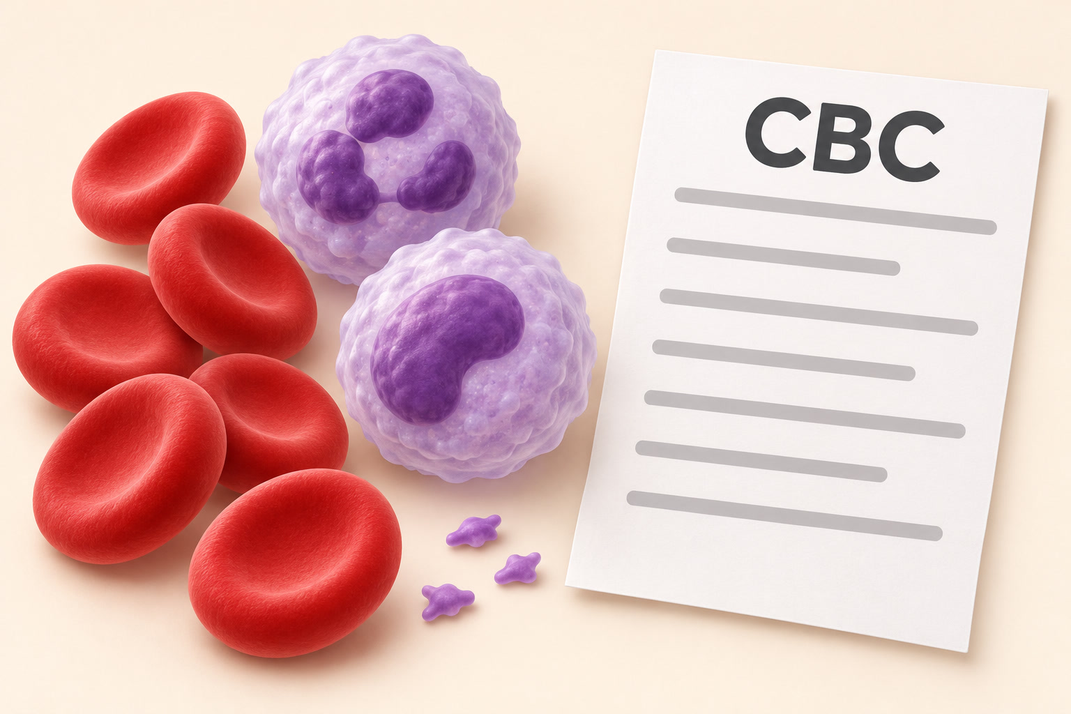
A complete blood count (CBC), also called a full blood count (FBC) or haemogram, looks at three main groups: red blood cells that carry oxygen, white blood cells involved in immune defence, and platelets that help stop bleeding. The extra abbreviations describe their size, number or haemoglobin content. A CBC with differential also breaks the white cells into their main types. MedlinePlus explains what a CBC includes.
Before you read any high or low flag
Use the reference range and units printed beside your own result. Ranges vary between laboratories and can differ with age, sex and pregnancy. Hydration, medicines, recent illness and other circumstances also matter.
An H, L or asterisk means the number falls outside that laboratory’s range. It does not, by itself, name a disease or tell you how urgent it is. A result inside the range does not rule out every problem either. Your symptoms, medical history, other results and changes over time give the numbers meaning. When possible, use the same laboratory to follow trends. How to understand laboratory results.
1. RBC: How many red blood cells are there?
RBC = red blood cell count. It counts red cells in a measured volume of blood, not in your entire body.
Think of red cells as oxygen-delivery vehicles. The RBC count tells you how many vehicles are present, but not how much oxygen-carrying protein each contains.
- A low count can occur with blood loss, nutritional deficiencies, kidney disease or problems with blood-cell production.
- A high count can reflect dehydration, living at altitude, low oxygen from some lung or sleep conditions, or increased red-cell production.
RBC is read together with haemoglobin and the red-cell indices. It is not a stand-alone measure of oxygen-carrying capacity. More about the RBC count.
2. Haemoglobin: The oxygen-carrying protein
Hb or Hgb = haemoglobin (also spelled hemoglobin). This is the protein inside red cells that carries oxygen. The result measures its concentration in whole blood, usually in g/dL or g/L.
Low haemoglobin, judged against the appropriate clinical threshold, indicates anaemia. It does not tell you the cause. Iron deficiency is one possibility, but blood loss, vitamin deficiencies, kidney disease, inflammation and other conditions can also contribute.
High haemoglobin can reflect concentrated blood from dehydration or increased red-cell production, among other causes. More is not automatically better.
Do not confuse Hb with HbA1c. HbA1c is a separate test used to assess average blood sugar. Haemoglobin testing explained.
3. Haematocrit: How much space do the red cells occupy?
Hct or PCV = haematocrit or packed cell volume. It describes the proportion of blood volume made up of red cells.
For example, an Hct of 40% means red cells occupy about 40% of the sample’s volume. This is an illustration, not a target value.
Hct often moves with haemoglobin. A low result can accompany anaemia or blood loss. A high result can occur with dehydration or increased red-cell production. The extra plasma, or liquid part of blood, during pregnancy can lower the proportion occupied by red cells. More about haematocrit.
4. MCV: The average size of a red cell
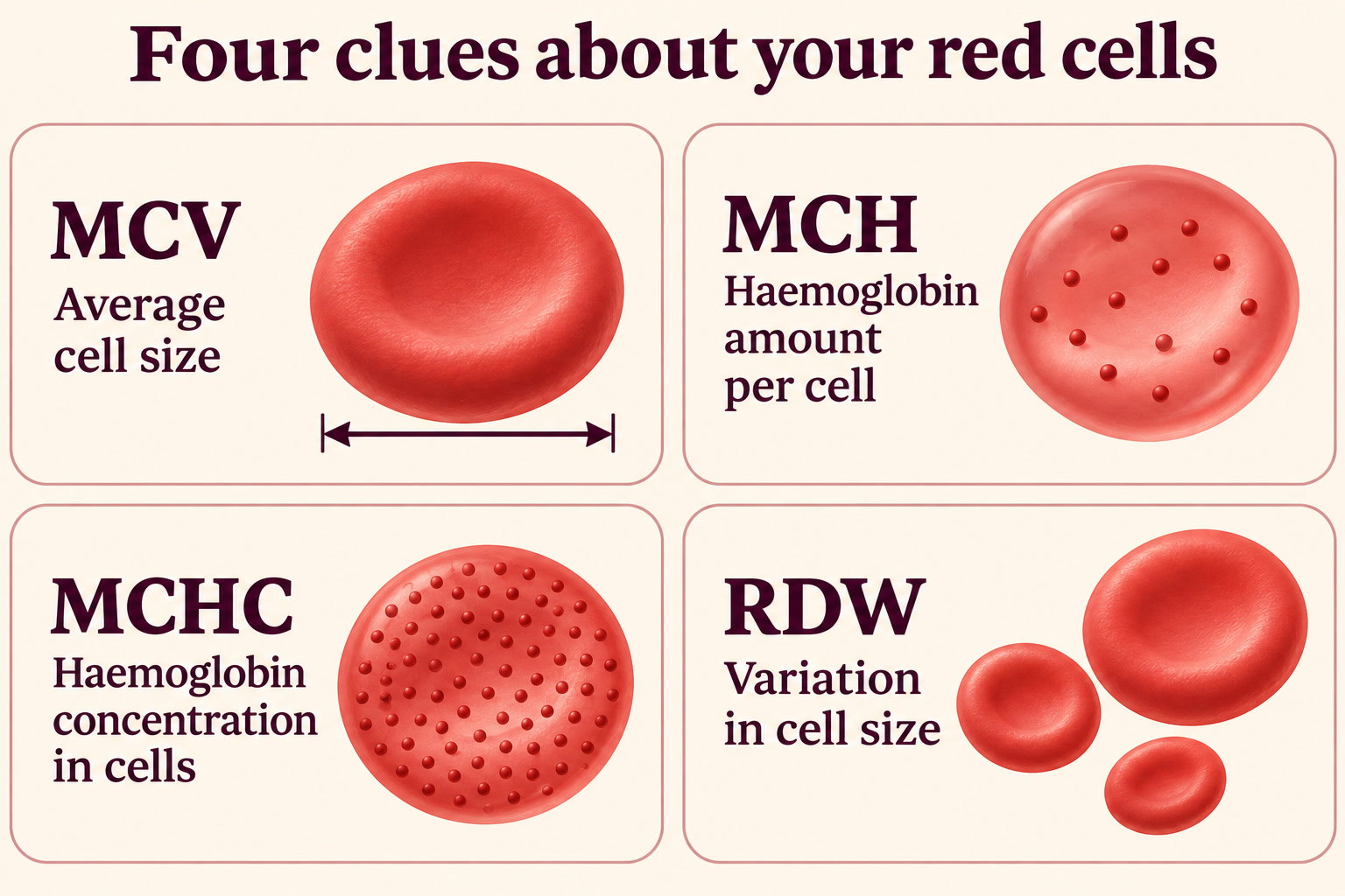
MCV = mean corpuscular volume. “Mean” means average, and “corpuscular” refers to a cell. Its usual unit is fL, a very small unit of volume.
- Low MCV: the cells are smaller on average, called microcytic. Iron deficiency and thalassaemia are possible explanations.
- High MCV: the cells are larger on average, called macrocytic. Possible explanations include vitamin B12 or folate deficiency, liver disease, alcohol effects, an underactive thyroid and some medicines.
- MCV within range: the average size is in range, called normocytic. You can still have anaemia, for example with kidney disease or blood loss.
MCV is an average. A mixture of small and large cells can produce an average that looks ordinary, which is one reason RDW and a blood film may help. MedlinePlus: MCV and NHS pathology: FBC interpretation.
5. MCH: How much haemoglobin is in each red cell?
MCH = mean corpuscular haemoglobin. It is the average amount of haemoglobin per red cell, usually measured in picograms (pg).
A smaller cell often contains less haemoglobin, so MCH commonly moves with MCV. Low MCH can occur with iron deficiency or thalassaemia. High MCH often accompanies larger red cells.
High MCH does not mean your iron stores are high. It describes haemoglobin per cell, not the amount of stored iron in your body. Red-cell indices explained.
6. MCHC: The concentration question
MCHC = mean corpuscular haemoglobin concentration. It describes how concentrated haemoglobin is within the red-cell volume, usually in g/dL or g/L.
Here is the distinction people often miss:
- MCV: How big is the average cell?
- MCH: How much haemoglobin does the average cell contain?
- MCHC: How much haemoglobin is present relative to the space the red cells occupy?
Imagine two containers of different sizes filled with the same-strength coloured liquid. The bigger container holds more liquid, but the concentration is unchanged. Similarly, a larger red cell may have a higher MCH without a higher MCHC.
Low MCHC means the haemoglobin is less concentrated. It can occur in iron-deficiency anaemia and thalassaemia. High MCHC can occur in conditions such as hereditary spherocytosis, which changes red-cell shape, and some conditions involving red-cell destruction. MedlinePlus: red-cell indices.
An unexpectedly high MCHC can also reflect interference with the measurement, such as red cells clumping in the sample. The laboratory may check the sample, repeat testing or examine a blood film. Do not use MCHC alone to diagnose a condition or decide to take iron. Review of unreliable automated CBC results.
7. RDW: Are the red cells similar in size?
RDW = red cell distribution width. It measures how much red-cell size varies, not how wide an individual cell is.
- High RDW: there is more variation in cell size. It can help investigate anaemia when read with haemoglobin, MCV and other tests.
- Normal RDW: the cells are fairly similar in size. They can still be uniformly small or large, so a normal RDW does not rule out anaemia.
- Low RDW: the sizes are very similar. On its own, this is usually not concerning.
Some reports show RDW-CV, usually as a percentage, and RDW-SD, usually in fL. They describe variation differently, so their numbers and reference ranges are not interchangeable. MedlinePlus: RDW and haematology reporting recommendations.
8. WBC or TLC: The total white-cell count
WBC = white blood cell count. TLC = total leucocyte count. Both terms refer to the total number of white cells in a measured volume of blood.
A high count may occur with infection, inflammation, physical stress, smoking, medicines such as steroids, or blood disorders. A low count may occur with some infections, medicines, immune conditions or bone-marrow problems.
WBC is not a simple “immunity score.” A high result does not automatically mean a bacterial infection, and a normal result does not exclude infection. The types of cells involved and your symptoms matter. White blood count explained.
9. The differential: Which white cells make up the total?
A differential leucocyte count (DLC) separates white cells into five main groups:
- Neutrophils: rapid responders that help fight infections, especially bacterial and fungal infections. Counts can rise with inflammation, stress or steroid treatment. A low absolute count is called neutropenia and can increase infection risk, depending on its severity and cause.
- Lymphocytes: include cells that make antibodies and cells that recognise or destroy infected cells. Counts can change with viral infections, medicines, immune conditions and blood disorders.
- Monocytes: help remove germs, damaged cells and debris. Higher counts can occur with some infections and inflammatory conditions.
- Eosinophils: are involved in allergic responses and defence against some parasites. A rise does not prove either an allergy or a parasitic infection.
- Basophils: a small group involved in allergic and inflammatory signalling. A persistent increase needs interpretation with the rest of the report.
These are roles and possible patterns, not a disease checklist. MedlinePlus: blood differential and differential result interpretation.
Absolute counts versus percentages: a crucial difference
A percentage tells you the share of white cells belonging to one type. An absolute count tells you how many of that type are present in a measured volume.
For example, if WBC is 8,000 cells/µL and lymphocytes are 25%, the absolute lymphocyte count is 2,000 cells/µL. If WBC is 4,000 and lymphocytes are 50%, the absolute count is still 2,000 cells/µL. These are arithmetic examples, not personal reference ranges.
So a flagged percentage may reflect a change in another cell type rather than a true increase or decrease in that cell’s number. Look for the absolute result, often marked ABS, #, ANC (absolute neutrophil count), ALC (absolute lymphocyte count) or AEC (absolute eosinophil count). Why proportions can change.
10. Platelets: The first responders to a blood-vessel leak
PLT = platelet count. Platelets are tiny cell fragments that help form the initial plug at an injury. They work with clotting proteins to stop bleeding.
- Low platelets, called thrombocytopenia, can increase bleeding risk. Causes include some infections, medicines, immune conditions and reduced production.
- High platelets, called thrombocytosis, can occur with inflammation, infection, iron deficiency or some bone-marrow disorders.
The cause, degree of change and symptoms matter. A platelet count alone does not show whether the platelets work normally, and it does not measure the entire clotting system. Platelet counts and function tests.
Sometimes platelets clump in the collection tube, producing a falsely low automated count. A repeat sample or blood film can clarify this. Do not assume an unexpected low count is just a sample problem; follow the clinician’s advice. CBC measurement caveats.
11. MPV: The average size of your platelets
MPV = mean platelet volume, usually reported in fL.
Newly released platelets are often larger, so a high MPV can be a clue that more young platelets are circulating. A low MPV means the average platelet size is smaller and can sometimes accompany reduced production.
Neither finding diagnoses the cause. MPV must be read with the platelet count, other results and your clinical situation. It is not a stand-alone test for clotting risk, heart disease or bone-marrow health. MPV explained.
Extra abbreviations some reports include
Not every laboratory reports all of these:
- Bands or IG (immature granulocytes): younger white-cell forms. Increases may accompany infection or other bone-marrow stress, but do not establish a diagnosis.
- NRBC (nucleated red blood cells): immature red cells that still contain a nucleus. They are different from reticulocytes. Their significance depends on age and clinical context; an unexpected finding in an adult needs medical review.
- PDW (platelet distribution width): how much platelet size varies.
- PCT (plateletcrit): the proportion of blood volume occupied by platelets. In this CBC context it is different from the separate infection-related test procalcitonin, which also uses the abbreviation PCT.
- P-LCR: the proportion of platelets classified as large by the analyser.
- Blood-film or morphology comments: observations about cell appearance under a microscope, such as size, shape, immature cells or clumping. These comments can be as important as the numbers.
Ask your clinician about unfamiliar flags rather than trying to interpret them alone. Report contents depend on the analyser and local laboratory practice. Sources: haematology reporting recommendations, platelet-index definitions, nucleated red cells on a blood film and NIH: blood-smear testing.
What a CBC does not measure
A CBC does not directly measure iron stores, vitamin B12, folate, blood sugar, kidney function or liver function. Depending on the pattern, a clinician may request ferritin and other iron studies, vitamin tests, kidney or liver tests, or a blood film. These are not automatically needed for everyone. RBC follow-up tests.
A reticulocyte count is often ordered separately rather than included in a routine CBC. Reticulocytes are young red cells. Their count helps show how the bone marrow is responding to the body’s need for red cells, including during anaemia or treatment. Reticulocytes explained.
A calmer way to read your report
- Check the date, units and laboratory reference range. Do not compare a child’s or pregnant person’s report with an unrelated adult chart.
- Read related results together. For red cells, look at haemoglobin, Hct, RBC, MCV, MCH, MCHC and RDW as a group.
- Compare with previous results. A new change can matter more than a stable personal pattern.
- Read the laboratory comments. “Platelet clumps” or a request to repeat the sample changes the interpretation.
- Ask what happens next. Does the result need monitoring, a repeat CBC, another test or treatment for an identified cause?
Avoid starting iron, vitamins, antibiotics or other treatment simply to “fix” a flagged number. The aim is to understand and address the cause.
When to seek medical help promptly
Do not wait for a report to become more abnormal if you feel seriously unwell. Seek emergency care for severe difficulty breathing, persistent or severe chest pain, collapse, or heavy bleeding that will not stop. NHS: breathing emergencies, chest pain and NIH: thrombocytopenia.
If you have known neutropenia or are receiving chemotherapy and develop a fever or feel acutely unwell, contact your treatment team or urgent medical service immediately and follow your treatment plan. NHS guidance on neutropenia.
If the laboratory or clinician calls a result critical, follow their urgent instructions even if you feel well. Urgency depends on the result and the person; one universal online cutoff cannot safely replace assessment.
The takeaway: RBC counts the cells. Haemoglobin measures the oxygen-carrying protein. Hct measures the space red cells occupy. MCV is size, MCH is amount, MCHC is concentration, and RDW is variation. White cells help with defence; platelets help stop bleeding. The whole pattern matters more than one isolated flag.
This article is for general education and is not a diagnosis or an individual treatment plan. Discuss your results with a qualified healthcare professional.
Sources and further reading
- MedlinePlus, U.S. National Library of Medicine: Complete blood count and understanding laboratory results.
- MedlinePlus: RBC count, haemoglobin and haematocrit.
- MedlinePlus: RBC indices, indices encyclopedia entry, MCV and RDW.
- MedlinePlus: WBC count, blood differential and differential interpretation.
- MedlinePlus: Platelet tests, MPV and reticulocyte count.
- North West London Pathology, NHS: Full blood count.
- National Heart, Lung, and Blood Institute, NIH: Platelet-disorder diagnosis and thrombocytopenia.
- StatPearls, hosted by NCBI Bookshelf: Normal and abnormal complete blood count with differential.
- Annals of Laboratory Medicine, hosted by PubMed Central: Unreliable automated complete blood count results: causes, recognition, and resolution.
- International Journal of Laboratory Hematology: Recommendations for standardising extended blood-count reporting units.
- Hematology, Transfusion and Cell Therapy, hosted by PubMed Central: Platelet indices and laboratory-specific reference intervals.
- Clinical Methods, hosted by NCBI Bookshelf: Peripheral blood smear.
- NHS: Shortness of breath, chest pain and NHS Highland neutropenia guidance.
#CBC #CompleteBloodCount #BloodReport #MCHC #MCV #MCH #RDW #Platelets #BloodHealth #HealthLiteracy #EndYourIllness
